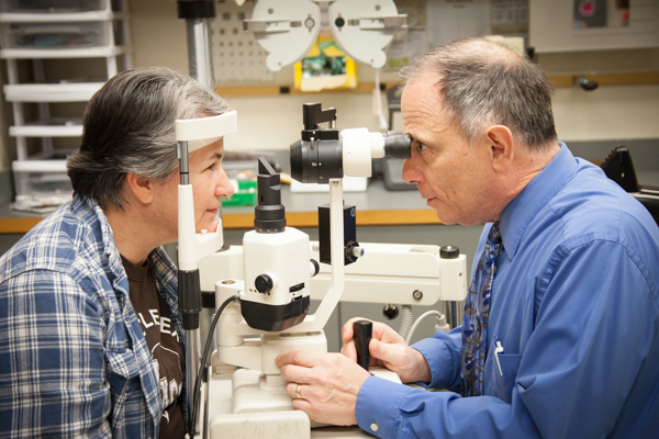
The Function of Advanced Diagnostic Devices in Identifying Eye Disorders
In the world of ophthalmology, the usage of innovative analysis devices has actually reinvented the early recognition and management of numerous eye conditions. From spotting refined adjustments in the optic nerve to checking the progression of retinal conditions, these innovations play a pivotal function in boosting the accuracy and performance of diagnosing eye problems. As the demand for accurate and prompt medical diagnoses proceeds to grow, the assimilation of advanced devices like optical comprehensibility tomography and visual area screening has ended up being indispensable in the realm of eye care. The complex interaction between technology and sensory practices not just clarifies detailed pathologies yet likewise opens up doors to customized therapy techniques.
Value of Very Early Medical Diagnosis
Very early diagnosis plays a pivotal duty in the effective management and therapy of eye disorders. By identifying eye conditions at an early phase, health care providers can use suitable therapy plans customized to the particular problem, eventually leading to better end results for clients.

Innovation for Spotting Glaucoma
Advanced diagnostic modern technologies play a critical duty in the early detection and surveillance of glaucoma, a leading root cause of irreparable blindness worldwide. One such innovation is optical comprehensibility tomography (OCT), which offers thorough cross-sectional photos of the retina, enabling the measurement of retinal nerve fiber layer thickness. This dimension is vital in analyzing damage triggered by glaucoma. Another sophisticated device is aesthetic field testing, which maps the level of sensitivity of a patient's visual area, aiding to spot any type of locations of vision loss characteristic of glaucoma. Furthermore, tonometry is made use of to measure intraocular stress, a major risk factor for glaucoma. This test is critical as elevated intraocular pressure can result in optic nerve damages. More recent technologies like the use of artificial intelligence algorithms in assessing imaging information are showing appealing outcomes in the early discovery of glaucoma. These sophisticated analysis tools make it possible for eye doctors to identify glaucoma in its early stages, enabling prompt intervention and much better monitoring of the condition to avoid vision loss.
Duty of Optical Coherence Tomography

OCT's ability to evaluate retinal nerve fiber layer density permits for precise and unbiased dimensions, assisting in the very early discovery of glaucoma even prior to visual field issues come to be noticeable. OCT modern technology permits longitudinal surveillance of architectural modifications over about his time, promoting individualized therapy strategies and timely treatments to help protect individuals' vision. The non-invasive nature of OCT imaging likewise makes it a recommended option for keeping an eye on glaucoma progression, as it can be repeated frequently without creating discomfort to the individual. Generally, OCT plays a vital duty in enhancing the analysis precision and monitoring of glaucoma, ultimately adding to much better results for individuals at danger of vision loss.
Enhancing Diagnosis With Visual Field Testing
A crucial part in detailed ophthalmic assessments, visual field testing plays an essential function in improving the analysis procedure for various eye problems. By examining the complete extent of a patient's visual field, this examination offers crucial information regarding the practical integrity of the whole aesthetic path, from the retina to the aesthetic cortex.
Aesthetic area testing is especially useful in the medical diagnosis and web monitoring of conditions such as glaucoma, optic nerve conditions, and various neurological conditions that can affect vision. Via quantitative measurements of peripheral and main vision, clinicians can spot subtle adjustments that may indicate the visibility or development of these problems, also before recognizable signs occur.
Additionally, aesthetic field testing permits the monitoring of treatment efficacy, assisting ophthalmologists tailor healing treatments to individual patients. eyecare near me. By tracking adjustments in aesthetic field efficiency gradually, medical care suppliers can make informed choices concerning readjusting drugs, advising medical treatments, or implementing other suitable steps to maintain or enhance a person's aesthetic function
Managing Macular Deterioration

Final Thought
In conclusion, advanced analysis tools play an important duty in recognizing eye conditions early on. Technologies such as Optical Comprehensibility Tomography and visual area testing have significantly boosted the precision and efficiency of identifying problems like glaucoma and macular deterioration. Early discovery permits timely treatment and monitoring of these conditions, ultimately bring about much better outcomes for clients. It is imperative for medical care specialists to remain upgraded on these advancements to give the ideal possible treatment for their people. eyecare near me.